
← Back to Atomic Structure contents
ATOMIC STRUCTURE · CHAPTER 10 OF 12
How is radiation used in medicine?
Radiation can reveal what is happening inside the body or damage unwanted tissue. Those two goals need different source choices, doses and ways of targeting the radiation.
By the end, you’ll be able to…
- explain how radioactive tracers help investigate organ function
- describe positron emission, annihilation and detection in PET
- explain why short-lived tracers need prompt production and delivery
- compare external radiotherapy, sealed implants and radioactive medicines
- evaluate benefits, dose and precautions for patients and staff.
Diagnosis or treatment?
For diagnosis, radiation is detected to gather information. The aim is a useful signal with exposure kept as low as reasonably achievable for that investigation.
For treatment, radiation delivers a controlled dose to damage unwanted cells. The aim is to treat the target while limiting damage to healthy tissue.
In nuclear medicine, a radioactive substance is introduced into the body. That differs from an ordinary X-ray image, where radiation comes from a machine outside the body.
A tracer follows a useful route
A radiopharmaceutical combines a radioactive isotope with a chemical form that travels to, or is taken up by, the tissue being investigated. A detector outside the body records radiation escaping from it.
A gamma-emitting tracer can provide information about organ function or where material accumulates. The chemical behaviour determines where it goes; the radioactive label makes its distribution detectable.
Material travels. Radiation escapes.
Watch the tracer travel in the bloodstream, collect in the organ and emit gamma radiation towards the detector. The detected pattern builds automatically.
* The movement, gamma rays and detector pattern are illustrative, not to scale. Real scans use many detection events and specialist image processing.
Choosing a radioactive tracer
- Suitable radiation: The isotope must emit radiation that can escape the body and be detected externally, such as gamma radiation.
- Suitable chemical form: The tracer must travel to the required organ or tissue so its uptake or movement can be investigated.
- Suitable activity: Enough activity to obtain useful information, without giving an unnecessarily high radiation dose.
- Suitable half-life: Long enough for preparation, delivery, uptake and scanning, but short enough for activity to decrease afterwards.
- Removal from the body: Biological removal and radioactive decay are different processes. Both affect how long radioactive material remains in the body.
* Technetium-99m is a widely used gamma-emitting isotope with a physical half-life of approximately 6 hours.
Which candidate fits an external gamma scan?
Assume all three chemical forms reach the organ of interest and have suitable starting activities. Preparation and scanning take several hours.
| Candidate | Emission | Half-life |
|---|---|---|
| A | Alpha | 6 h |
| B | Gamma | 6 h |
| C | Gamma | 600 years |
* Hypothetical candidates illustrate selection principles, not prescribing decisions.
PET: detect the photons produced after positron emission
Positron emission tomography (PET) uses a positron-emitting tracer. A positron has the same mass as an electron but a positive charge.
After travelling a short distance, the positron encounters an electron. They annihilate, producing two gamma photons travelling in approximately opposite directions. A ring of detectors records pairs arriving almost simultaneously.
The scanner detects the gamma photons, not the positron directly. Many detected pairs allow a computer to reconstruct the tracer distribution.
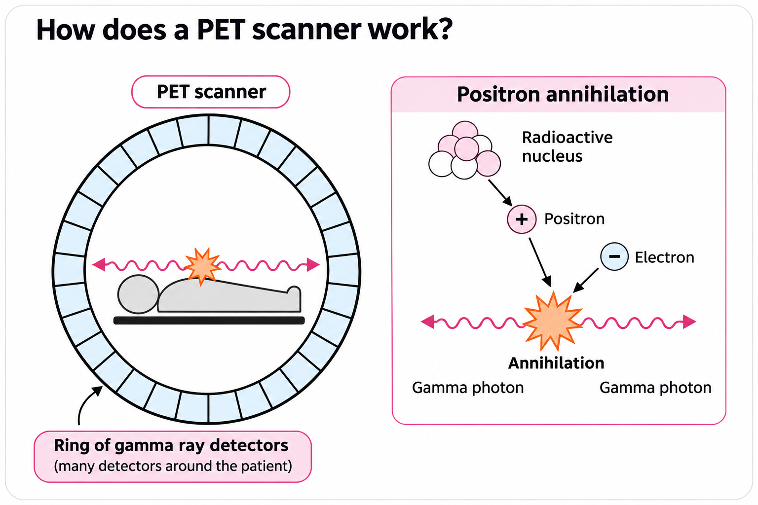
Positron emission
A radioactive tracer inside the body emits a positron, which travels a short distance before meeting an electron.
*A positron has the same mass as an electron but a positive charge.
Annihilation
The positron and electron annihilate, producing two gamma photons that travel in approximately opposite directions.
Detection
Detectors around the patient record photon pairs arriving almost simultaneously. Many detections help a computer build an image of the tracer's distribution.
Why are PET isotopes often produced near hospitals?
PET isotopes often have short half-lives. This example shows how activity can decrease during the time taken to deliver a tracer.
Activity decreases with time
From production to hospital
A shorter journey means less activity is lost before the scan.
* The 20-minute half-life and activity values are hypothetical. Actual PET isotopes have different half-lives, and some are transported from regional production centres.
Radiotherapy: damage unwanted tissue
Radiotherapy uses ionising radiation to damage DNA in unwanted cells, including cancer cells, so they die or cannot continue dividing. Healthy cells can also be damaged, so treatment planning aims to deliver the prescribed dose to the target while limiting exposure elsewhere.
External radiotherapy directs beams into the body from outside. High-energy X-rays are commonly produced by a machine; gamma rays from a radioactive source can also be used. Penetrating radiation can reach a target beneath the skin.
Why use more than one direction?
Compare one X-ray beam with several beam directions. See how the beams reach the same area from different angles.
Internal treatment: place or carry the source near the target
Some treatments place a radioactive source close to the tumour, while others use radioactive medicine taken up by particular tissues. Compare these methods below.
Where is the radiation source?
Select each treatment to find out where the radiation comes from.
Where is the source?
Outside the body
A machine directs high-energy X-rays towards the tumour. No radioactive material is placed inside the patient.
Where is the source?
Inside or close to the tumour
A small sealed radioactive source delivers radiation locally. It may be removed after treatment or left in place.
Where is the source?
Inside the body
A radioactive substance is given to the patient and is taken up by particular tissues.
Protecting patients and staff
During diagnosis, enough radioactive tracer must be used to produce a useful image, while keeping radiation exposure as low as reasonably practicable.
During treatment, radiation is carefully directed towards the tumour to minimise damage to surrounding healthy tissue.
Medical staff reduce their exposure by limiting the time spent near radiation sources, increasing their distance and using appropriate shielding.
Three worked examples
These hypothetical data practise physics calculations; they are not clinical dose or prescribing instructions.
Calculate remaining tracer activity:
A tracer starts at 160 activity units and has a 2 h half-life. Find its activity after 6 h.
Number of half-lives = time ÷ half-life
6 ÷ 2 = 3; 160 → 80 → 40 → 20
Activity = 20 units
Calculate corrected count rate:
A detector records 3600 counts in 60 s. Background is 5 counts/s.
Corrected rate = (counts ÷ time) − background rate
Corrected rate = (3600 ÷ 60) − 5
Corrected rate = 55 counts/s
Calculate dose in millisieverts:
A supplied hypothetical exposure is 0.0035 Sv.
Dose in mSv = dose in Sv × 1000
Dose = 0.0035 × 1000
Dose = 3.5 mSv
Does the smallest dose always give the best investigation?
Two hypothetical protocols investigate the same organ. A uses a lower dose but may not produce a usable image; B uses a higher dose and reliably provides the required information. No numerical risk or patient details are given.
Check your understanding
Choose one answer for each question. Feedback appears underneath.
“PET and radiotherapy both use radiation, so they must work in the same way. If a PET isotope has a very short half-life, it is always the best choice.” Explain the mistakes.
Distinguish diagnosis from treatment. Include what PET detects, delivery time and healthy-tissue exposure.
Reveal the answer
PET gathers diagnostic information from paired gamma detections following positron–electron annihilation. Radiotherapy aims to deliver a controlled dose to damage unwanted cells.
A short physical half-life reduces lingering activity, but it must still allow production, delivery, uptake and a useful scan. An isotope that decays too quickly may not provide enough activity at the right time.
Both applications can expose healthy tissue, so the radiation type, chemical targeting, activity and procedure must be chosen for the task. The expected benefit must justify the exposure; a short half-life alone does not make every source suitable.
CHAPTER 10
Chapter summary
- Diagnostic tracers reveal distribution or function; their chemical form directs where they travel.
- A suitable gamma-emitting tracer can be detected outside the body.
- PET uses positron emitters; annihilation with an electron produces two approximately opposite gamma photons.
- Many paired detections allow reconstruction; one pair alone does not locate an exact point.
- Short-lived PET isotopes need timely production and delivery.
- Radiotherapy damages unwanted tissue using external beams or internal sources.
- Targeting limits healthy-tissue exposure but does not eliminate it.
- Patient benefit, useful signal or target dose, and precautions for patients and staff must all be considered.